Anatomy Of Upper Thigh And Hip - 3d Human Upper Leg Anatomy Or Anatomical And Muscle Set Stock Illustration Illustration Of Foot Health 198223856 : This vein, as well as the deep veins.
byLuis Wall•
0
Anatomy Of Upper Thigh And Hip - 3d Human Upper Leg Anatomy Or Anatomical And Muscle Set Stock Illustration Illustration Of Foot Health 198223856 : This vein, as well as the deep veins.. The upper part of the thigh bone consists of the femoral head, femoral neck, and greater and lesser trochanters. Each pelvic girdle consists of a hip bone (coxal bone, innominate bone), which articulates with the head of a femur. Hip surgeon dr guillaume dumont offers hip pain treatments in columbia, sc. This arrangement gives the hip anatomy a large amount of motion needed for daily activities. Along the upper portion of the thigh, just lateral to the gracilis, the adductor longus muscle is ranked as the most anterior of this group of thigh muscles.
The patient lies supine with the hip and knee flexed and the hip externally rotated into the frog leg position. Use the mouse scroll wheel to move the images up and down alternatively use the tiny arrows (>>) on both side of the image to move the images. Quadriceps, a group of four. Knowing the anatomy of your hip can help you understand the source of any hip pain. for detailed anatomy of pelvic bones, read anatomy of hip bone.
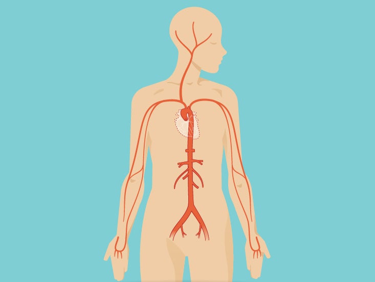
Thigh Anatomy Diagram Pictures Body Maps from post.healthline.com This arrangement gives the hip anatomy a large amount of motion needed for daily activities. The thigh is the area between the hip and the knee joint. The median cubital vein (a common site site for venepuncture) in the antecubital fossa of the arm. Hip and knee pain and hip and shoulder pain are. Knowing the anatomy of your hip can help you understand the source of any hip pain. The patient lies supine with the hip and knee flexed and the hip externally rotated into the frog leg position. The hip's unique anatomy enables it to be both extremely strong and amazingly flexible, so it can bear weight and allow for a wide range of movement. Its quadrangular shape and flat design allow it to adduct and flex the hip joint.
The upper part of the thigh bone consists of the femoral head, femoral.
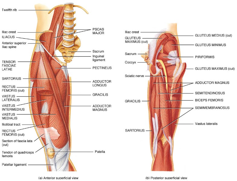
Diarthrodial joint with its inherent stability dictated primarily by its osseous components/articulations. It's restricted by contact of the thigh with all the abdomen and adduction is restricted by contact. Medial condyle of tibia nerve supply: On the anterior side, the most prominent of the muscles are the sartorius muscle and the four the four muscle of the quadriceps all extend the lower leg, and the rectus femoris additionally can flex the thigh at the hip. The muscles also require a lot of blood flow, which provides oxygen and nourishment, especially when you're physically active. This webpage presents the anatomical structures found on hip mri. Superficial fascia.—the superficial fascia forms a continuous layer over the whole of the thigh; The uppermost of the medial thigh muscles is the pectineus muscle. Tibial part of the sciatic nerve action: Pelvis, perineum, hip, and upper thigh. All of the anatomical parts of the hip work together to enable various movements. The thigh is the area between the hip and the knee joint. Hip and knee pain and hip and shoulder pain are.
The femur, the hip bone (subdivided into ilium. During hip replacement surgery, your surgeon removes the upper part of your thigh bone, including the femoral head (ball of the hip joint) and a part the upper part of the thigh bone is then exposed, and a series of tools called broaches are introduced one at a time to prepare your thigh bone for a metal. The median cubital vein (a common site site for venepuncture) in the antecubital fossa of the arm. It's restricted by contact of the thigh with all the abdomen and adduction is restricted by contact. He also serves the communities of charleston, sc and augusta, ga.
Tensor Fasciae Latae Muscle Hip And Upper Thigh Pain Hip Stiffness The Wellness Digest from thewellnessdigest.com In vertebrate anatomy, hip (or coxa in medical terminology) refers to either an anatomical region or a joint. Anatomy hip, thigh and leg muscles. Hip surgeon dr guillaume dumont offers hip pain treatments in columbia, sc. While the thigh muscles will be slip into the anterior, medial and posterior groups. The joints and muscles of the hips and thighs need nervous input so they can do what your brain wants them to do. Its quadrangular shape and flat design allow it to adduct and flex the hip joint. Mri of upper leg (femur). This webpage presents the anatomical structures found on thigh mri.
431).—at the upper and medial part of the thigh, a little below the medial end of the inguinal ligament, is a large.
Anatomy ▶ lower limb ▶ bones and cartilages ▶ hip joint. All of the anatomical parts of the hip work together to enable various movements. Mri of upper leg (femur). The femur, the hip bone (subdivided into ilium. Jew anatomy atlases, the anatomy atlases logo, and a digital library of anatomy information are all trademarks of michael p. The adductor muscle on the inner thigh; The femur or thigh bone is one of the longest bones in the human body. This webpage presents the anatomical structures found on thigh mri. The muscles also require a lot of blood flow, which provides oxygen and nourishment, especially when you're physically active. Like the forearm, the upper leg, or thigh, has a dense arrangement of many muscles. Upper part of the ischial tuberosity insertion: The anatomical areas found on the upper limb can serve as key landmarks to help us find important anatomical structures such as finding one of the superficial veins: Chief flexor of knee weak.
Tibial part of the sciatic nerve action: The following nerves serve the gluteal and. B, muscles of the anterior thigh compartment. Upper part of the ischial tuberosity insertion: This mri hip joint axial cross sectional anatomy tool is absolutely free to use.
Hip Area Anatomy Anatomy Drawing Diagram from boneandspine.com Tibial part of the sciatic nerve action: It functions to adduct the thigh and to flex. The thigh is the area between the hip and the knee joint. The uppermost of the medial thigh muscles is the pectineus muscle. Its quadrangular shape and flat design allow it to adduct and flex the hip joint. During hip replacement surgery, your surgeon removes the upper part of your thigh bone, including the femoral head (ball of the hip joint) and a part the upper part of the thigh bone is then exposed, and a series of tools called broaches are introduced one at a time to prepare your thigh bone for a metal. The upper part of the thigh bone consists of the femoral head, femoral. The adductor muscle on the inner thigh;
Use the mouse scroll wheel to move the images up and down alternatively use the tiny arrows (>>) on both side of the image to move the images.
for detailed anatomy of pelvic bones, read anatomy of hip bone. A, anterior and posterior views show the hip joint ligaments. Jew anatomy atlases, the anatomy atlases logo, and a digital library of anatomy information are all trademarks of michael p. The femur or thigh bone is one of the longest bones in the human body. The following nerves serve the gluteal and. Use the mouse scroll wheel to move the images up and down alternatively use the tiny arrows (>>) on both side of the image to move the images. The adductor muscle on the inner thigh; Superficial fascia.—the superficial fascia forms a continuous layer over the whole of the thigh; The muscles also require a lot of blood flow, which provides oxygen and nourishment, especially when you're physically active. Several muscles cross the front of the hip and create hip flexion, pulling the thigh and trunk toward both muscles cross the floor of the pelvis, emerge at the outer edges of the pubic bones, and finally insert on the inner upper femur (thighbone). Muscles of the hips and thighs. Pelvis, perineum, hip, and upper thigh. 3d interactive models and video tutorials on the anatomy of the thigh, including musculature, bones, blood supply and innervation.
The hip muscles are going to be slip into hip muscles and gluteal muscles upper thigh anatomy. Unlike the shoulder girdle, the pelvic girdle is firmly integrated into the axial skeleton: